|
|
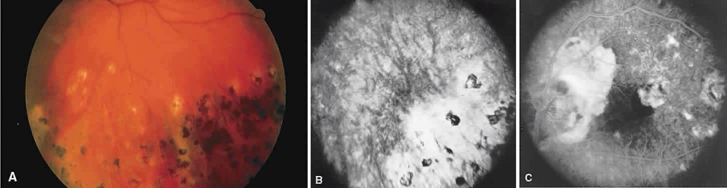
| Fig. 4. Sympathetic ophthalmia. A. Areas of pigmentation and small areas of focal atrophy. B. Fluorescein angiogram showing blocked fluorescein corresponding to the areas of pigmentary disturbance with areas of hyperfluorescence representing old areas of inflammation. C. Blocked fluorescence due to subretinal blood in the juxtafovea with associated choroidal neovascularization. |