|
|
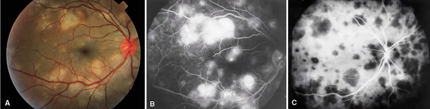
| Fig. 3. Vogt-Koyanagi-Harada syndrome. A. Fundus photograph of the left eye showing multifocal serous retinal detachment. B. Late fluorescein angiogram showing pooling of dye in the areas of serous detachment. C. Indocyanine angiography showing areas of blocked fluorescence corresponding to serous detachment. More areas of hypofluorescence are noted than those observed on fluorescein angiogram. |