|
|
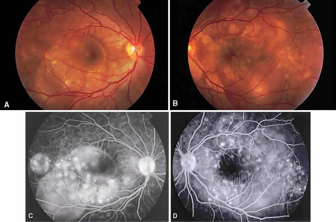
| Fig. 2. Vogt-Koyanagi-Harada syndrome. (A) Fundus photograph of the right eye and (B) the left eye showing bullous retinal detachments. (C) Fluorescein angiogram showing multiple hyperfluorescent leaks at the level of the retinal pigment epithelium. (D) Multiple coalescing leaks and fluorescein staining of the subretinal fluid. Large areas of hyperfluorescence involving the entire right posterior pole due to pooling of dye beneath the subretinal space. |