|
|
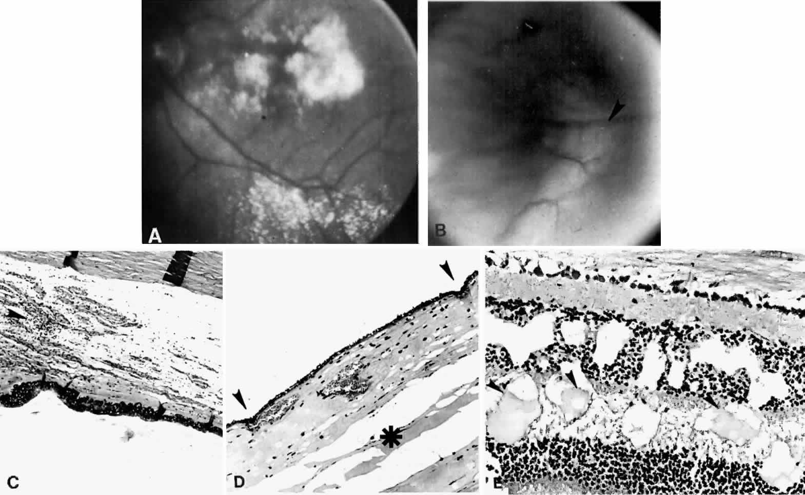
| Fig. 21. A. Retinal exudates situated inferotemporally in the left eye of a 46-year-old woman with idiopathic ciliochoroidal effusion mistaken for a malignant melanoma and associated histopathologically with mild uveitis. Exudate in the macular area had a yellowish-orange appearance. B. Appearance of “solid-detachment” ciliochoroidal effusion with a meridionally oriented fold (arrowhead). C. Pars plana area with faintly stained proteinaceous material separating the smooth muscle fibers and tangentially oriented fibers associated with a mild lymphocytic infiltration (arrowhead) (H & E, × 50). D. Effusion of the choroid (asterisk) near the equator by a more densely stained proteinaceous material with a very light scattering of lymphocytes. Circumferentially oriented folds (arrowheads) involving the retinal pigment epithelium and inner aspect of the choroid are evident (H & E, × 135). E. Edema and cystic changes in the retina below the fovea. Most cysts in the inner nuclear layer have no staining material present, but those in the outer plexiform layer have a lightly staining proteinaceous material (arrowheads) (H & E, × 215). |