|
|
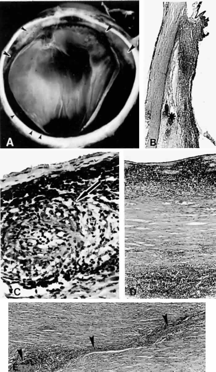
| Fig. 15. A spontaneous ciliochoroidal effusion was mistaken for a malignant melanoma in a 61-year-old woman who presented with pain, blurred vision, keratitic precipitates, and aqueous cells and flare. A. Gross appearance of the ciliochoroidal effusion (large arrowheads). Small whitish inflammatory nodules are present in the choroid (small arrowheads). The apparent retinal detachment is artifact. B. An extensive ciliochoroidal effusion (asterisk) with a moderately intense chronic inflammatory cellular infiltration. (H E, × 20). C. The choroid viewed posteriorly, showing occlusive granulomatous vasculitis (arrow) and an intense lymphocytic infiltration crowding the choriocapillaris. The retinal pigment epithelium is intact (H E, × 290). D. Intense lymphocytic infiltration of choroid, inner scleral lamellae, and episclera (H E, × 55). E. Diffuse scleritis viewed posteriorly, showing perineural lymphocytic infiltration in the episclera and within a scleral canal (arrowheads) (H E, × 55). |