|
|
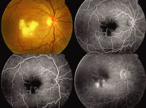
| Fig. 31. Colored fundus photography and fluorescein angiography of the right eye of a male patient who presented with sudden drop of vision following high fever 3 days previously. In the ophthalmoscopic view of the right eye there is extensive retinal whitening in the macular area (necrotic retina) with macular edema. Angiography reveals early blocked fluorescence corresponding to retinal necrosis seen in the central fundus. In the late arteriovenous phase of the angiogram, there is complete occlusion of the some perifoveal capillary arterioles, which end abruptly. During the very late phase of the angiogram, the picture of blocked fluorescence by the retinal necrosis is still seen centrally, however, there is late staining of some macular capillary arterioles. Late dye leakage from these damaged perifoveal capillaries with marked perivascular staining of these capillaries is indicative of perivasculitis. This fundus picture of acute necrotizing retinitis with perivasculitis and multiple arteriolar occlusion gives the typical picture of Rift Valley Fever retinitis. (Courtesy of M Moussa, Tanta University, Egypt.) |