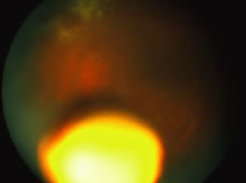
Fig. 21.
Colored fundus photo showing the intravitreal ganciclovir implant after insertion. An area of active cytomegalovirus (CMV) retinitis is seen in the upper portion of the illustration.