|
|
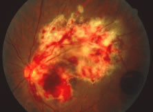
| Fig. 13. A patient with hemorrhagic variant of cytomegalovirus (CMV) retinitis that affected the posterior pole and optic nerve. Hemorrhages are most often intraretinal, and white areas correspond histologically to intracellular and extracellular edema and necrosis of the neurosensory retina. Eyes with extensive optic nerve involvement secondary to retinal involvement may still retain good central vision unless the papillomacular bundle is involved. When retinitis starts in the optic nerve head, the prognosis for vision is poor. |