|
|
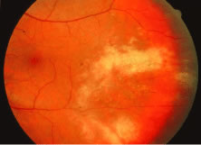
| Fig. 12. A patient with a relatively nonhemorrhagic variant of cytomegalovirus retinitis. Note at the center of the lesion (oldest area of infection) the retina is atrophic and pigmentary changes can be seen. Peripheral to it the retina is edematous, and this is the area of active infection. |