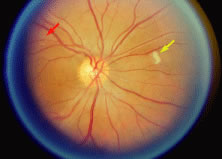
Fig. 8.
A patient with cytomegalovirus (CMV) lesion superotemporal to the optic nerve (
red arrow
) and cotton-wool lesion (
yellow arrow
) superonasal to the optic nerve. The CMV lesion is granular and deep.