|
|
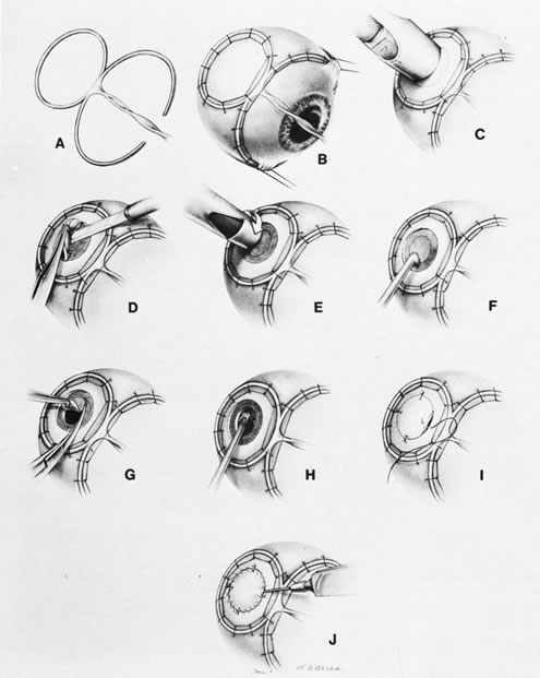
| Fig. 16. A. Peyman eye biopsy basket is placed. B. Eye basket is sutured to the globe. C. Trephine marks the site. D. Partial-thickness scleral flap is made. E. Full-thickness scleral trephine is done. F. Surface diathermy is performed. G. Sclerochorioretinal biopsy specimen is taken. H. Vitrectomy is done. I. Sclera is sutured in place. J. Ocular volume is reconstituted by air or fluid injection, and a vitrectomy is performed as in Figure 13D. |