|
|
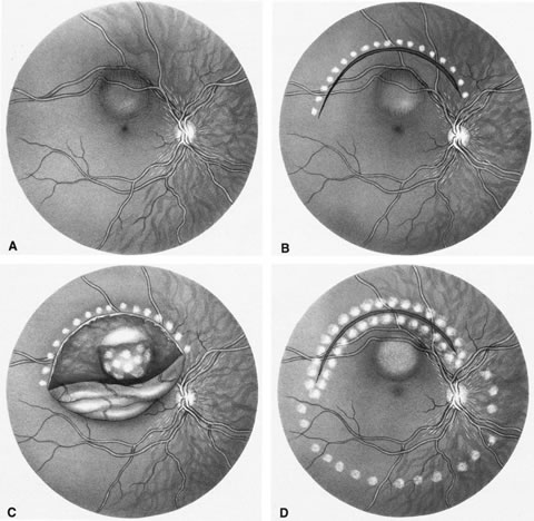
| Fig. 15. A. Uveal melanoma located in the posterior pole within 2 disc diameters of the optic nerve and within 0.5 disc diameter of the fovea. B. Following endodiathermy, a 180-degree arcuate retinotomy is performed, ensuring a 1 disc–diameter tumor-free margin. C. The retinal flap is dissected, and the entire tumor exposed. Argon endolaser is applied both to tumor surface and free margins. Tumor and free margins are then dissected from surrounding tissue bed and removed. D. Following tumor removal and complete vitrectomy, the retinal flap is repositioned and an air-fluid exchange performed. Endolaser is applied to the retinotomy edges and the posterior pole. (Peyman GA, Nelson NC Jr, Paris CL, et al: Internal choroidectomy of posterior uveal melanomas under a retinal flap. Int Ophthalmol 16:439, 1992. Reprinted by permission of Kluwer Academic Publishers.) |