|
|
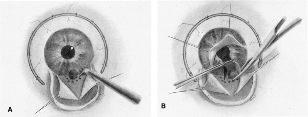
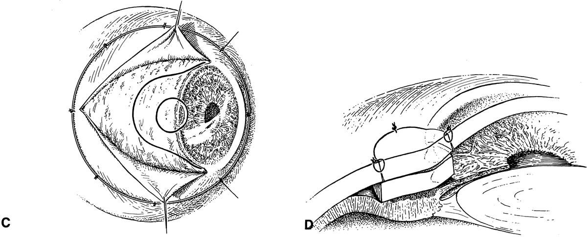
| Fig. 5. A. A T-shaped incision is made over the tumor to expose the mass outlined by transillumination. Diathermy is placed around the tumor-free margins. B. The pupillary margin is grasped and the iris incised radially to the iris root. The tumor is excised by grasping the pupil margin with Vannas scissors. C. Curved scleral-limbal incision with trephine of Müller. The area over the tumor is trephined through the cornea and sclera. D. The corneoscleral button is depressed, and the scleral graft is sutured in place. The tumor is then excised with the corneoscleral tissue. |