|
|
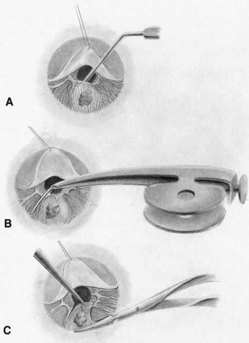
| Fig. 1. A. Corneal-scleral wound is opened, and a viscoelastic substance is placed under the iris to protect the lens. B. The cornea is retracted by an assistant, and iris scissors are used to cut the tumor radially with clear margins. C. Iris section is grasped and excision along the iris root is performed. |