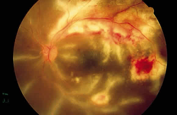
Fig. 13.
Fundus photograph of the right eye of a patient with cytomegalovirus retinitis and extensive “frosted branch angiitis.” The active cytomegalovirus retinitis is temporal to the fovea and along the superotemporal arcade.