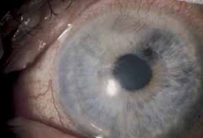
Fig. 17
Sectoral iris atrophy, status post–herpes zoster ophthalmicus. Note the moth-eaten appearance of iris in sectoral area, underlying a corresponding area of sclerokeratitis and limbal vascularization.