|
|
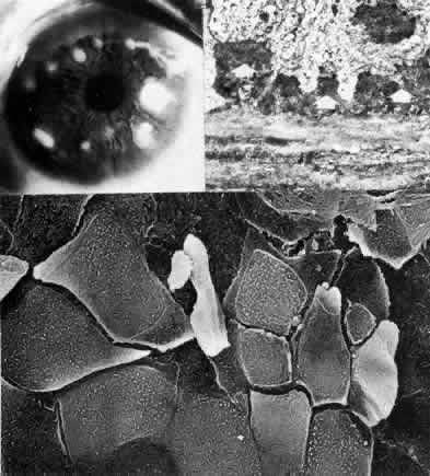
| Fig. 29. Salzmann's nodular degeneration. Top left. Clinical photograph emphasizes numerous discrete elevated opacities in the anterior midperipheral stroma. Top right. Transmission electron micrograph of corneal epithelium over opacity reveals marked irregular thickening of basement membrane material (arrows) overlying collagenous pannus (× 7500). Bottom. Scanning electron micrograph of epithelial surface covering a nodular deposit shows extreme disorganization and breakdown of epithelial mosaic with extensive desquamation and separation of adjacent cells (× 1200). (Bottom, courtesy of Diane Van Horn, PhD) |