|
|
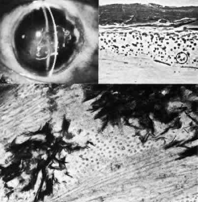
| Fig. 28. Band keratopathy. Top left. In a 59-year-old woman with congenital luetic interstitial keratitis, band keratopathy has resulted in epithelial erosion with persistent central defect. Top right. Phase-contrast microscopy discloses irregular corneal epithelium with a myriad of small, densely staining spherules (circled) within Bowman's layer (paraphenylenediamine, × 1000). Bottom. Transmission electron micrograph resolves fine crystalline characteristic and extreme electron density of calcium or hydroxyapatite particles (× 70,000). |