|
|
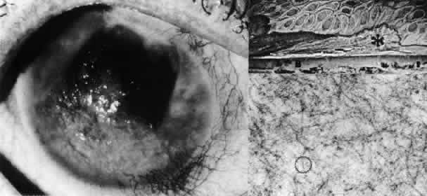
| Fig. 26. Amyloid degeneration. Left. In a patient with long-standing stromal and epithelial edema due to Fuchs' dystrophy, superficial irregular opacities consist of amyloid material. Top right. Phase-contrast microscopy shows masses of amyloid material (*) intervening between the epithelium and Bowman's layer (B) (paraphenylenediamine, × 750). Bottom right. Transmission electron micrograph of amyloid deposit shows typical ultrastructural characteristics of 8- to 10-nm diameter, banded (circled) fibrils in random aggregates (× 60,000). |