|
|
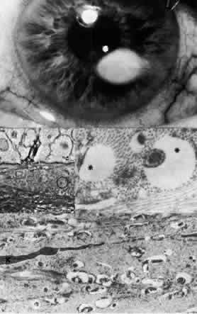
| Fig. 25. Lipid degeneration. Top. Clinical photograph shows opaque lipid deposit with central vessel. Middle left. Phase-contrast microscopy includes numerous fine extracellular deposits (circled) within Bowman's layer and anterior stroma (paraphenylenediamine, × 800). Bottom. Transmission electron micrograph of anterior stroma illustrates globular lipids among collagen fibrils without disruption or other abnormality of keratocytes (K) (× 12,000). Middle right. At higher magnification, lipid deposits of approximately 1μm diameter have characteristics of saturated neutral fats (*) (× 40,000). (Top. Grayson M: Diseases of the Cornea, p 194. St. Louis, CV Mosby, 1979) |