|
|
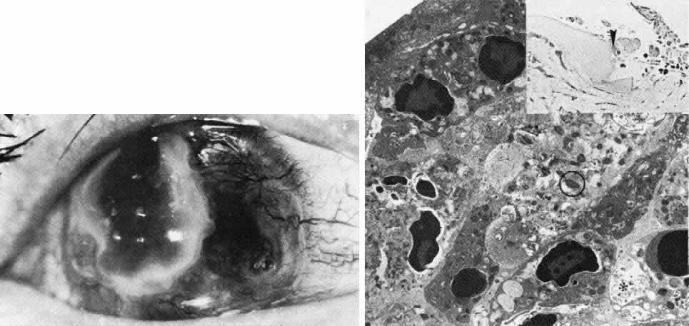
| Fig. 24. Mooren's ulcer. Top. Clinical photograph in an advanced bilateral case shows extensive loss of peripheral stroma that has vascularized with only central island of full-thickness stroma remaining. Middle right. Phase-contrast micrograph of stroma at margin of ulcerating area includes abrupt termination of Bowman's layer (at arrowhead) with numerous acute inflammatory cells shown at right (paraphenylenediamine, × 800). Bottom. Transmission electron micrograph of area in previous illustration resolves multiple intrastromal inflammatory cells as polymorphonuclear leukocytes actively engaged in degranulation and phagocytosis (*). The stroma is extensively degraded in areas where enzyme-containing leukocyte granules (circled) are present (× 7500). |