|
|
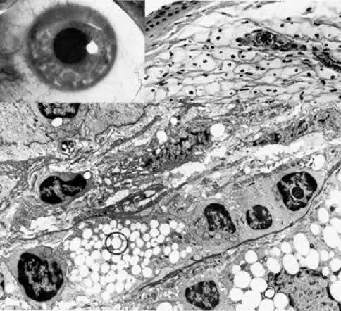
| Fig. 23. Terrien's marginal degeneration. Top left. Clinical photograph of a patient with extensive peripheral thinning of superior stroma shows vascularization of involved stroma with lipid deposition at advancing edge. Top right. Light microscopy reveals numerous foamy histiocytic cells and blood vessels within the anterior stroma (hematoxylin-eosin, × 300). Bottom. Transmission electron micrograph shows several histiocytic cells laden with neutral lipid inclusions (circled). Several reactive fibroblasts and chronic inflammatory cells are also seen (× 5000). |