|
|
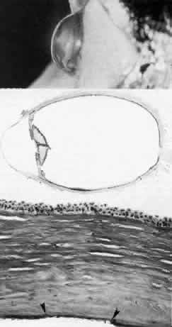
| Fig. 21. Keratoglobus. Top. Clinical photograph of acquired keratoglobus in a 65-year-old man shows bulging globoid contour of corneas that were clear except for small stromal opacities in the temporal midperiphery. Middle. Horizontal pupil-optic nerve section of this eye shows bulging cornea and deep anterior chamber. The entire cornea is approximately one-third normal thickness, except in extreme periphery nasally and temporally (hematoxylin-eosin, × 4). Bottom. Light photomicrograph of central cornea shows old rupture of Descemet's membrane (between arrowheads) with subsequent deposition of new thinner Descemet's membrane by regenerated endothelium (hematoxylin-eosin, × 400). (Green WR: Keratoglobus. Am J Ophthalmol 77:393, 1974) |