|
|
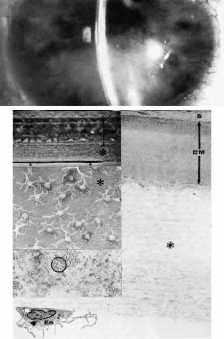
| Fig. 19. Chandler's syndrome. Top. Clinical appearance of cornea in a 37-year-old woman with unilateral stromal edema, peripheral anterior synechiae, iris atrophy, and glaucoma. Upper middle left. Phase-contrast microscopy shows abnormal thick acellular posterior collagen layer (*) between Descemet's membrane (DM, bracketed) and the discontinuous endothelium (arrowheads) (paraphenylenediamine, × 1400). Center middle left. Scanning electron micrograph of posterior corneal surface shows remaining endothelial cells to have attenuated dendritiform configuration. Large areas of Descemet's membrane and posterior collagen layer (*) are exposed (× 250). Bottom right. Transmission electron micrograph corresponding to phase-contrast photograph (upper middle left) reveals relatively normal stroma (S) and Descemet's membrane (DM) with posterior layer (*) composed of loosely arrayed fibrillar collagen and basement membrane material. A single attenuated endothelial cell (En) is evident at the far left (× 6000). Lower middle left. Transmission electron micrograph detail of posterior collagen layer resolves bundles of fine filaments (circled) plus fibrillar collagen with segment long-spacing (SLS)-banding (× 50,000). (Top, courtesy of R Meyer, MD) |