|
|
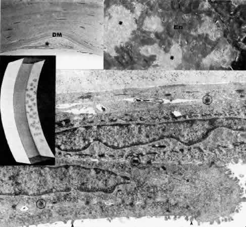
| Fig. 18. Posterior polymorphous dystrophy. Middle left. Slit lamp drawing emphasizes typical vesicular lesions at the level of Descemet's membrane. Top left. Phase-contrast microscopy of posterior stroma and Descemet's membrane (DM) demonstrates the focal deposition of posterior collagenous material (*), presumably corresponding to vesicular lesions (paraphenylenediamine. × 600). Top right. Scanning electron micrograph demonstrates the extreme polymorphous configuration of endothelial cells (En) with intervening areas of exposed Descemet's membrane (*) consistent with the corneal edema of this keratoplasty specimen (× 200). Bottom. Transmission electron micrograph shows endothelial cells to have transformed into epithelial-appearing tissue, as multiple cell layers have numerous interconnecting desmosomes (circled) and individual cells show increased keratofibrils and microvillous surface projections (arrowheads) (× 19,000). |