|
|
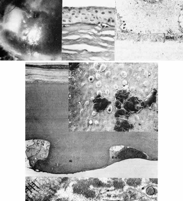
| Fig. 17. Late hereditary endothelial dystrophy (Fuchs). Top left. Clinical photograph illustrates epithelial bullae, scarring, and neovascularization, resulting from long-standing stromal edema. Top middle. Light microscopy demonstrates intraepithelial edema, thickening of the basement membrane, subepithelial bullae (*) and fibrocellular pannus with adjacent break in Bowman's layer (hematoxylin-eosin, × 350). Top right. Transmission electron micrograph of basal epithelial cells and Bowman's layer shows multilaminar basement membrane complexes (BM, the sequel of chronic epithelial edema (× 5000). Middle left. Transmission electron micrograph of posterior cornea shows unremarkable stroma and anterior Descemet's membrane, but remarkable thickening of posterior Descemet's membrane to 12 mm with additional superimposition of large guttata (G). The remaining endothelial cells (En) are severely degenerated and attenuated (× 5000). Middle right. By scanning electron microscopy, the comparable picture of disjointed, attenuated endothelium (En) and numerous exposed guttata (*) is apparent. Note the fibrous feltwork quality of the abnormal posterior Descemet's membrane (× 300). Bottom. High-magnification trans mission electron micrograph of guttata resolves its composition of fine filaments (circled), multiple segments of basement membrane material (*), and collagen in long-spacing configuration (arrowheads) (× 50,000). (Top left. Grayson M: Diseases of the Cornea, p 242. St. Louis, CV Mosby, 1979) |