|
|
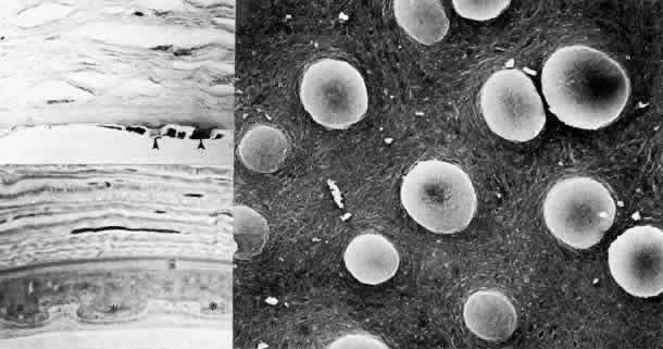
| Fig. 16. Corneal guttata. Top left. By light microscopy, excrescences (arrowheads) of Descemet's membrane are evident with loss of endothelial cells (hematoxylin-eosin, × 250). Bottom left. Phase-contrast microscopy resolves thickened Descemet's membrane with individual guttata (.) having been covered posteriorly by additional collagenous material (paraphenylenediamine, × 1000). Right. Scanning electron micrograph of posterior corneal surface with endothelium removed shows numerous mushroom-shaped excrescences projecting from the surface of Descemet's membrane (× 1000). (Right, courtesy of Diane Van Horn, PhD) |