|
|
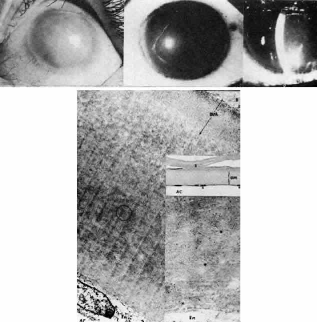
| Fig. 15. Congenital hereditary endothelial dystrophy. Top left. Clinical photograph of eye of a 14-year-old male with severe form of the dystrophy shows diffuse ground-glass stromal opacification. Top middle. In a mildly affected 20-year-old female, the cornea has moderate diffuse haze and visual acuity is 20/200. Top right. On slit lamp biomicroscopy, diffuse edematous thickening of the corneal stroma is evident in same patient as top middle photograph. Middle right. Light microscopy of a case with uniformly thickened (approximately 35μm) Descemet's membrane (DM) covered posteriorly by extremely attenuated endothelial cells (arrowheads). S, posterior stroma; AC, anterior chamber (hematoxylin-eosin, × 600). Bottom left. Transmission electron micrograph of same case as middle right micrograph reveals anterior portion of Descemet's membrane (DM) to have normal thickness and banded structure. The markedly thickened (approximately 20 μm) posterior layer exhibits both 55 nm and 110 nm banding (circled) interspersed with homogeneous material. En, endothelial cell; AC, anterior chamber; S, posterior stroma (× 9200). Bottom right. At higher magnification, the abnormal posterior zone is seen to consist of multiple laminations of basement membrane-like material (*) and fine filaments. En, endothelial (× 42,000). |