|
|
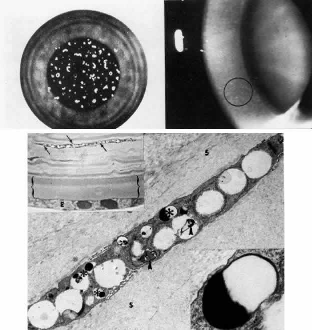
| Fig. 14. Grayson-Wilbrandt dystrophy. Top left. Artistic representation of large polymorphous deposits in the pre-Descemet's membrane area, having comma-shaped, circular, linear, filiform, and dotlike configurations. Top right. Slit lamp photograph shows discrete pleomorphic opacities (circled) with clarity of the intervening stroma. Bottom. Upper left inset demonstrates by phase-contrast microscopy the refractile vacuolar inclusions (arrows) within a deep keratocyte. Descemet's membrane (bracketed) is uniformly normal and endothelial cells (E) are artifactiously vacuolated (toluidine blue, × 1000). Main figure is a transmission electron micrograph of a keratocyte filled with vacuoles having clear to fibrillogranular material, pleomorphic substances (arrowheads). and dark, electron-dense bodies (*). The surrounding stroma (S) is normal (× 12,000). Lower right inset by higher magnification transmission electron micrograph shows homogeneous electron density and limiting membrane of the intracytoplasmic inclusion indicated by the large asterisk in the main figure (× 40,000). (Curran RE, Kenyon KR, Green WR: Pre-Descemet's membrane corneal dystrophy. Am J Ophthalmol 77:711, 1974) |