|
|
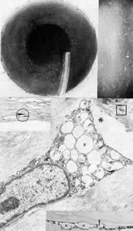
| Fig. 13. Fleck dystrophy (Francois-Neetens). Top left. Artistic representation of discrete, flattened white stromal flecks shows comma, wreath, or dot configuration. Top right. Retroillumination slit lamp photograph demonstrates similar configuration of small, white, granular opacities throughout the stroma. Bottom. Lower right inset is phase-contrast micrograph of a severely affected keratocyte showing foamy cytoplasm with large clear vacuoles (*) and small refractile inclusions (arrowheads) (paraphenylenediamine × 1400). Upper left inset illustrates positive staining for acid mucopolysaccharide limited to a swollen keratocyte (circled) (colloidal iron, × 500). Main figure is transmission electron micrograph of a markedly vacuolated keratocyte filled with fibrillogranular (F) or lipid (L) substances. There are no extracellular abnormalities except an accumulation of the fine granular material (,) and occasional foci of long-spacing collagen (square) (× 12,000). (Nicholson DH, Green WR, Cross HE et al: A clinical and histopathological study of Francois Neetens speckled corneal dystrophy. Am J Ophthalmol 83:554, 1977) |