|
|
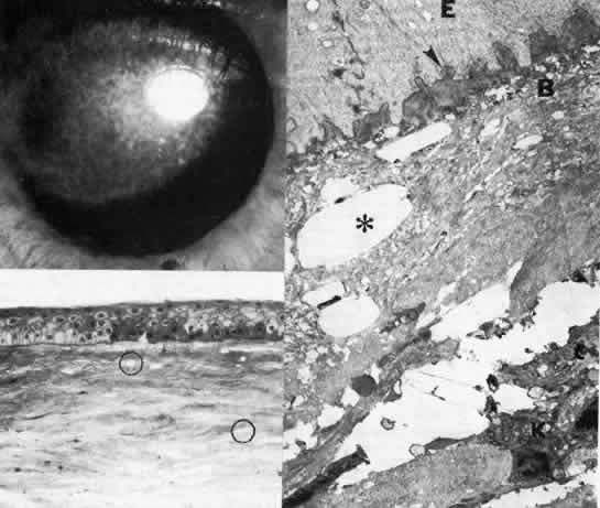
| Fig. 12. Central crystalline dystrophy (Schnyder). Top left. Clinical appearance of eye of 20-year-old woman includes ovoid crystalline deposit with clear surrounding stroma and without arcus lipidis. Visual acuity is 20/40. Bottom left. Light microscopy of cornea demonstrates epithelial irregularity and numerous crystalline profiles (circled) in Bowman's layer and stroma (toluidine blue, × 350). Right. Transmission electron micrograph demonstrates basal epithelium (E) with thickened basement membrane complexes (arrowhead), disorganized Bowman's layer (B), and polygonal crystalline profiles (*) typical of cholesterol. The keratocyte (K) is unremarkable (× 10,400). (Gipson I: Schnyder's crystalline dystrophy. Trans Am Ophthalmol Soc 76: 184, 1978) |