|
|
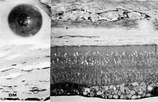
| Fig. 11. Macular corneal dystrophy. Top left. Clinical appearance of cornea features diffuse haze extending to the limbus with superimposed, dense gray-white spots. Bottom left. Light photomicrograph of posterior cornea shows endothelial cells staining intensely positive for acid mucopolysaccharide. Guttate excrescences (*) of Descemet's membrane (DM) are frequent. The stroma also shows positive staining for acid mucopolysaccharide both diffusely extracellularly and intensely within keratocytes (circled) (colloidal iron × 500). Right. Transmission electron micrograph discloses typical fibrillary granular deposits within keratocytes (K), throughout the posterior layer of Descemet's membrane, and within the endothelial cells (En). The anterior banded region of Descemet's membrane (bracketed) is not affected (× 3500). |