|
|
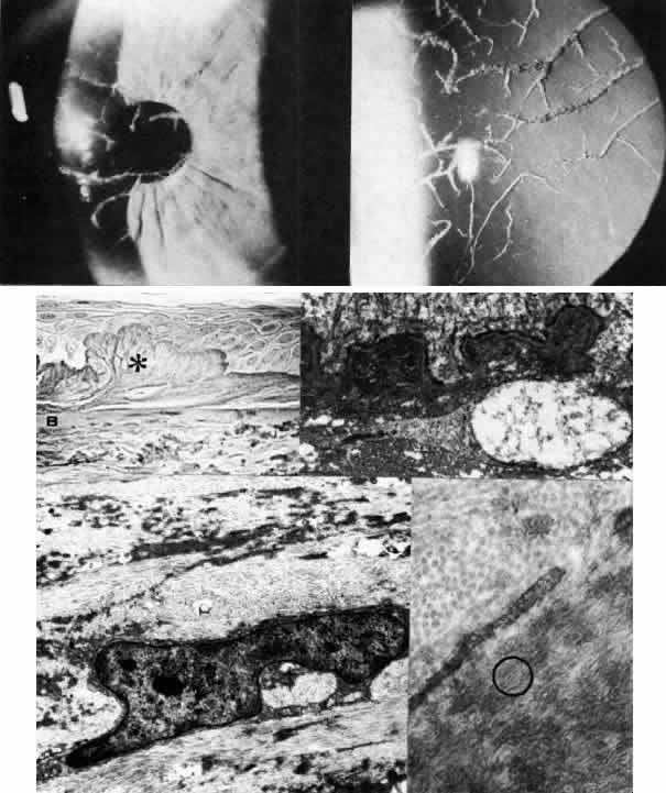
| Fig. 10. Lattice corneal dystrophy. Top. Slit lamp photograph demonstrates pathognomonic branching lattice figures throughout the stroma. Middle left. Phase-contrast photomicrograph shows subepithelial accumulations of fibrillar amyloid deposits (*) causing distortion of epithelial contour. B, Bowman's layer (paraphenylenediamine, × 800). Middle right. Transmission electron micrograph of basement membrane complexes reveals basement membrane irregularity and discontinuity resulting from underlying amyloid fibrils (× 21,000). Bottom left. Transmission electron micrograph of stroma shows normal collagen fibrils and keratocytes with electron-dense material abnormally dispersed extracellularly (× 16,000). Bottom right. High-magnification transmission electron micrograph resolves lattice material as masses of fine, 8- to 10-nm diameter amyloid fibrils (circled below) in comparison with larger-size stromal collagen fibrils (above) (× 75,000). (Slit lamp photographs courtesy of WJ Stark, MD) |