|
|
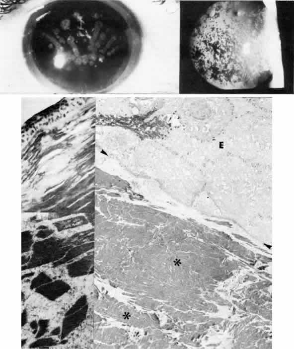
| Fig. 9. Granular corneal dystrophy. Top left. Discrete, large opacities predominantly affect the central stroma. Top right. Retroillumination emphasizes the optical clarity of intervening stroma between granular opacities. Middle left. Light microscopy of hyaline deposits is accentuated with Masson trichrome stain (× 250). Right. Transmission electron micrograph shows relatively normal epithelium (E) and basement membrane (arrowheads) anterior to large electron-dense deposits (*) within Bowman's layer and stroma (× 4,500). Bottom left. Higher magnification transmission electron micrograph of granular deposits shows characteristic rod-shaped paracrystalline structure ( × 50,000). (Top right, Slit lamp photograph courtesy of Lawrence Hirst, MD) |