|
|
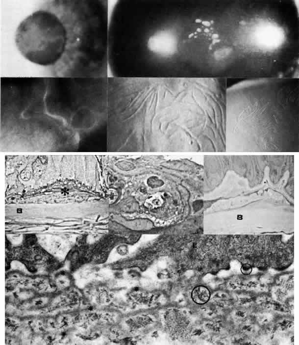
| Fig. 6. Map-dot-fingerprint dystrophy. Top left. Clinical photograph of a 37-year-old man with non-traumatic erosions shows characteristics of map dystrophy with superficial geographic haze interrupted by clear areas. Top right. In the dot form of Cogan's mycrocystic dystrophy, superficial, opaque cysts are evident within the epithelium. Upper middle. Three variants of fingerprint dystrophy show subepithelial ridges, particularly enhanced by retroillumination. Lower middle left. Phase-con-trast microscopy of map dystrophy shows fibrous tissue (*) interposed between epithelium and Bowman's layer (B) (paraphenylenediamine, × 1000). Lower middle center. Phase-contrast microscopy of dot dystrophy shows an intraepithelial pseudocyst evolving from disintegration of desquamating cells (paraphenylenediamine, × 1200). Lower middle right. Phase-contrast micrograph of fingerprint dystrophy illustrates fingerlike intraepithelial extensions of aberrant fibrocellular material anterior to the normal-appearing Bowman's layer (B) (paraphenylenediamine, × 800). Bottom. Transmission electron micrograph in these disorders consistently finds multiple laminations of basement membrane material (*) with reduced hemidesmosomes (small circle) and increased anchoring fibrils (large circle) beneath epithelium (E) (× 40,000). (Upper middle, slit lamp photographs courtesy of Lawrence Hirst, MD) |