|
|
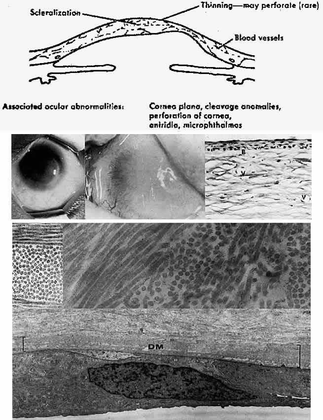
| Fig. 4. Sclerocornea. Schematic drawing of ocular features Top left. In a minimally affected patient with additional findings of ptosis, strabismus, and hearing loss, only the peripheral cornea is opacified. Top center. In this advanced case with chromosomal translocation and multiple congenital abnormalities, the entire cornea is sclerified and the fine vascular arcades extend centrally from the conjunctiva and sclera. Top right. Light micrograph of anterior cornea shows edematous disorganization of epithelium, fragmentation of Bowman's membrane (B), and interstitial vascularization (V) (hematoxylin-eosin, × 200). Middle left. Transmission electron micrograph of normal human corneal stroma is shown for comparative purposes. Note uniform 240- to 260-nm collagen fibril diameter (× 50,000). Middle right. Transmission electron micrograph of sclerocornea at same magnification shows disorganized array of collagen fibrils that measure as much as three times normal diameter (× 50,000). Bottom. Transmission electron micrograph of posterior cornea shows abnormal Descemet's membrane of less than 1μm thickness (DM, bracketed) and attenuated endothelial cells (× 10,500). (Schematic. Grayson M: Diseases of the Cornea, p 32. St. Louis, CV Mosby, 1979; Top center and right. Rodrigues MM, Calhoun J, Weinreb S: Sclerocornea with an unbalanced translocation [17p, 10q]. Am J Ophthalmol 78:49, 1974) |