|
|
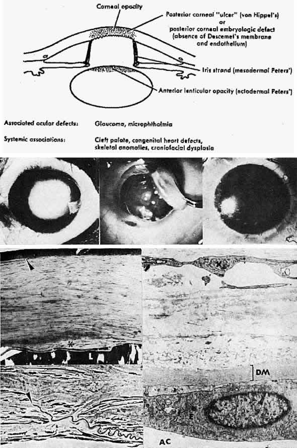
| Fig. 3. Peters' anomaly. Schematic drawing of ocular features. Top left. Clinical photo of typical bilateral Peters' anomaly with large, dense central leukomata, which was successfully treated by penetrating keratoplasty with optical iridectomy of the fellow eye. Top center. Intraoperative photo demonstrates adhesion of the lens to the posterior cornea as a corneal button (grasped with forceps) is trephined. Top right. Successful penetrating keratoplasty of a patient with bilateral Peters' anomaly. Bottom, upper left. Survey light photomicrograph of a corneal button from the case illustrated clinically (top center) shows termination of Bowman's membrane (at arrowhead) corresponding to the area of adhesion between the posterior cornea and lens (L). Descemet's membrane, present peripherally, terminates centrally in a layer of retrocorneal fibrous tissue (*) interposed between the lens and stroma. Direct contact between the retrocorneal fibrous tissue and continuous lens capsule is evident (paraphenylenediamine, phase contrast, × 60). Bottom, lower left. Phase-contrast micrograph of posterior cornea adjacent to a central stromal defect shows termination (at arrowhead) of undulating Descemet's membrane between the stroma and retrocorneal fibrous tissue (paraphenylenediamine, × 400). Bottom right. Transmission electron micrograph of posterior cornea shows attenuated keratocytes (K) with phagocytic contents, disorganized posterior stromal lamellae, and markedly thin and multilaminar Descemet's membrane (DM) with attenuated but continuous endothelium (E). AC, anterior chamber. (x 7000) (Schematic. Grayson M: Diseases of the Cornea, p 29. St. Louis, CV Mosby, 1979) |