|
|
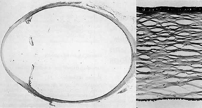
| Fig. 1. Megalocornea. Left. Light microscopy of a 62-year-old man with corneal diameters of 13 mm shows in pupil-optic nerve section an enlarged anterior segment with no abnormalities (except beveled scar of cataract incision and surgical aphakia) (hematoxylin-eosin, × 3). Right. Central section of the cornea demonstrates all layers to be normal except for some thinning of the epithelium (hematoxylin-eosin, × 165). (Greene WR: Md State Med J, July 1974) |