|
|
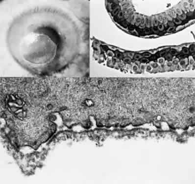
| Fig. 31. Diabetic corneal erosion. Top left, Epithelial defect in a diabetic patient after vitrectomy. Top right, Phase-contrast photomicrograph demonstrating intact epithelial sheet removed during vitreoretinal surgery because of loose adherence. (Paraphenylenediamine, × 400.) Bottom, Electron micrograph of basal cell surface from epithelial scraping showing intact basement membrane attached to epithelial cells. (× 25,000.) (Courtesy of Dr. Kenneth Kenyon) |