|
|
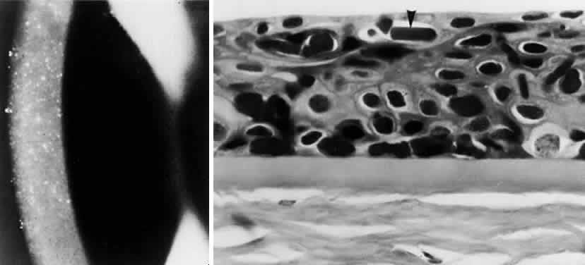
| Fig. 10. Multiple myeloma. Top, Crystals in cornea. Bottom, Crystalline deposits (arrow) in epithelium. (Masson trichrome, × 680.) (Klintworth GK, Bredehoeft SJ, Reed JW: Analysis of corneal crystalline deposits in multiple myeloma. Am J Ophthalmol 86:303, 1978) |