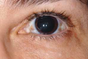
Fig. 11
Oculodermal melanocytosis or nevus of Ota. Note the scleral pigmentation as well as dermal pigmentation of the upper and lower lateral eyelid.