|
|
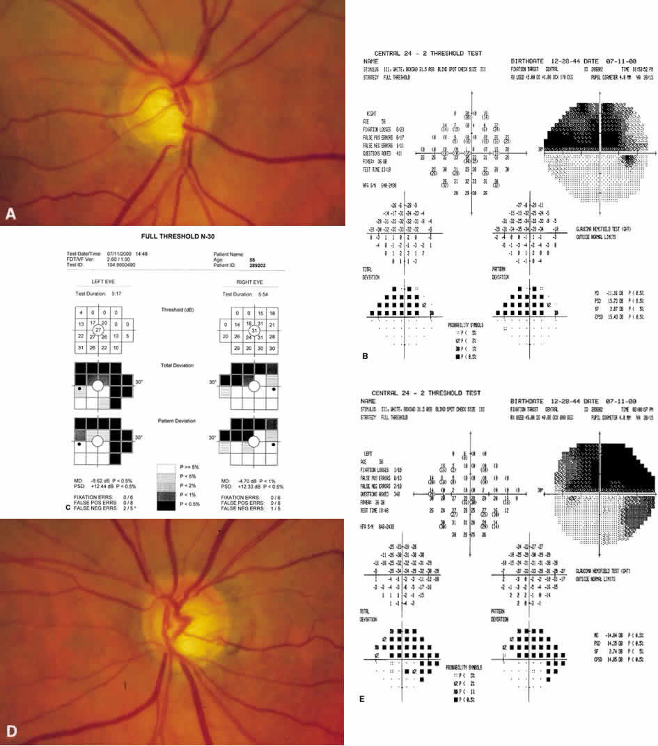
| Fig. 25. A. Fundus photograph of right optic nerve. Note an inferior notch. Corresponding superior visual field defect on Humphrey Field Analyzer (HFA; B) andfrequency doubling technology (FDT; C). D. Left optic nerve with inferior notch and superior thinning. E. Visual fields of the left eye show a larger defect in the superior than inferior hemifield on both HFA and FDT. |