|
|
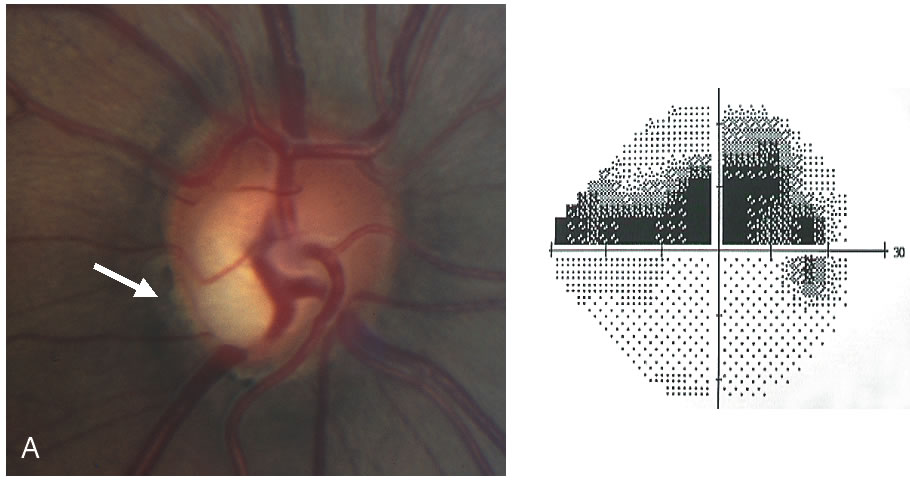
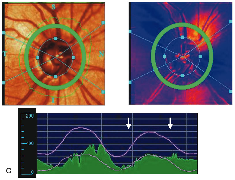
| Fig. 22. Comparison of imaging techniques: glaucomatous eye. A. Marked thinning of the inferior neuroretinal rim is seen in this glaucomatous eye with a notch at the 7 o'clock position. There is a corresponding superior arcuate defect on visual field testing. B. The optic disc is classified as outside normal limits by Heidelberg Retina Tomograph (HRT; Heidelberg Engineering, Heidelberg, Germany) with Moorfield's classification system. C. Attenuation of retardation and a flattening of the double hump curve in the inferotemporal region are noted on scanning laser polarimetry (SLP). D. The optical coherence tomography (OCT) shows a reduction of the nerve fiber layer in this region to 40 μm. |