
|
|
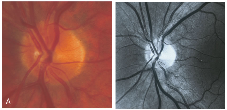
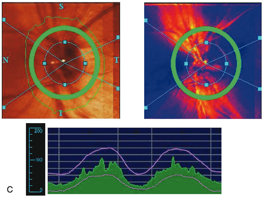
| Fig. 21. Comparison of imaging techniques: healthy eye. A. A healthy appearing neuroretinal rim is seen. B. The optic disc is classified as within normal limits by Heidelberg Retina Tomograph (HRT; Heidelberg Engineering, Heidelberg, Germany) with Moorfield's classification system. C. A typical double hump curve is seen on scanning laser polarimetry (SLP). D. The anterior and posterior highly reflecting layers, representing the retinal nerve fiber layer (RNFL) and retinal pigment epithelium (RPE), can be seen on optical coherence tomography (OCT). |