|
|
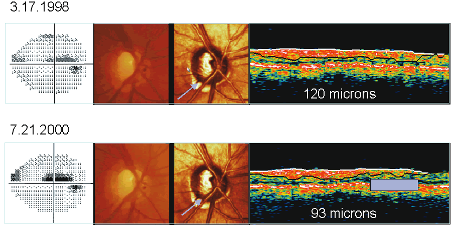
| Fig. 20. Optical coherence tomography: monitoring change. In this example, a baseline and follow-up image of an uncontrolled glaucomatous eye is shown. Thinning of the retinal nerve fiber layer (RNFL) in the inferotemporal quadrant is seen with worsening of the corresponding superior arcuate visual field defect. |