|
|
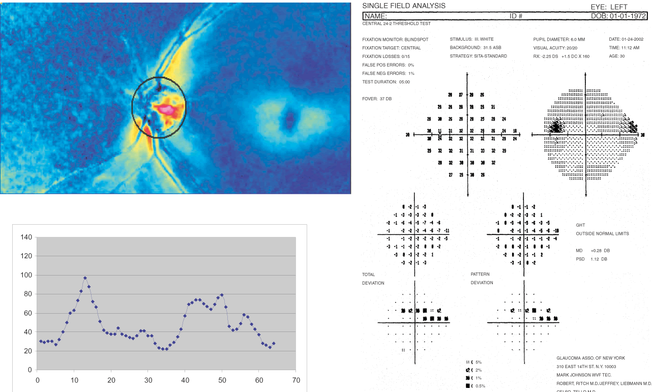
| Fig. 13. Scanning laser polarimetry: glaucomatous eye. In this example of a glaucomatous eye, thinning of the superotemporal neuroretinal rim with a corresponding inferonasal visual field defect is seen. Retardation is markedly attenuated and the double hump curve is significantly depressed in the superotemporal quadrants. |