|
|
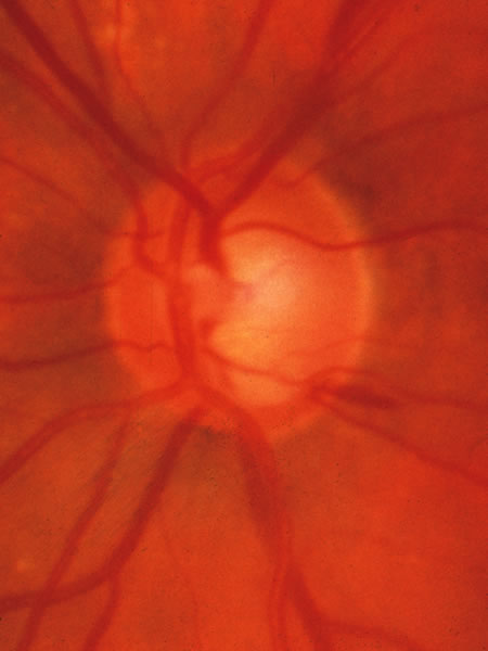
| Fig. 32. An obviously glaucomatous disc with particular loss of tissue in the inferior and superior sectors. A splinter hemorrhage among the axons crossing the disc margin is present at the 4:30 meridian. Typical glaucomatous hemorrhages are elongated and usually extend from the disc tissue across the disc margin somewhat into retina. They may overlie an adjacent peripapillary zone of choroidal or pigment epithelial atrophy, but usually have at least one end touching the disc margin. Nonglaucomatous hemorrhages are usually obvious in context of diabetic retinopathy, retinal vein occlusion, papillitis, etc. |