|
|
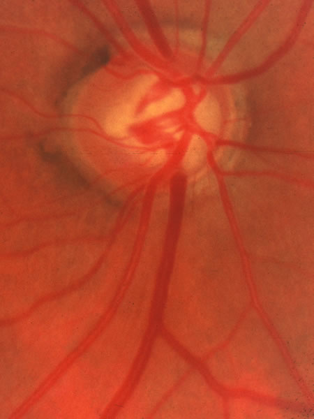
| Fig. 28. Glaucomatous disc with cupping to the nasal side. A thin remnant of neuroretinal tissue remains just inside the white stripe of the scleral lip in the lower nasal region. Under some lighting conditions the adjacent β zone of atrophy may be mistaken for intact neuroretinal tissue. The field loss extending temporally from the blind spot may be overlook if perimetry covers only the central 24 degrees and the significance of one or two abnormal spots temporal to the blind spot is dismissed as artifact. |