|
|
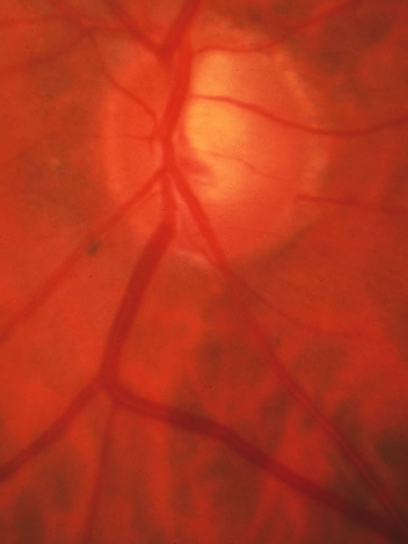
| Fig. 25. Glaucomatous disc with excavation fairly localized to the superior sector. In this case the vessels penetrate the lamina cribrosa in the inferior half of the disc. The deviation of the small vessel near the upper pole marks the surface configuration. The upward expansion of the pale base of the cup nearly to the disc margin is a distinctive sign that certainly signifies abnormality, but can be missed by indirect ophthalmoscopy if the strong lighting imparts a pink color to the region and the subtle small vessel deviation is not noticed. |