|
|
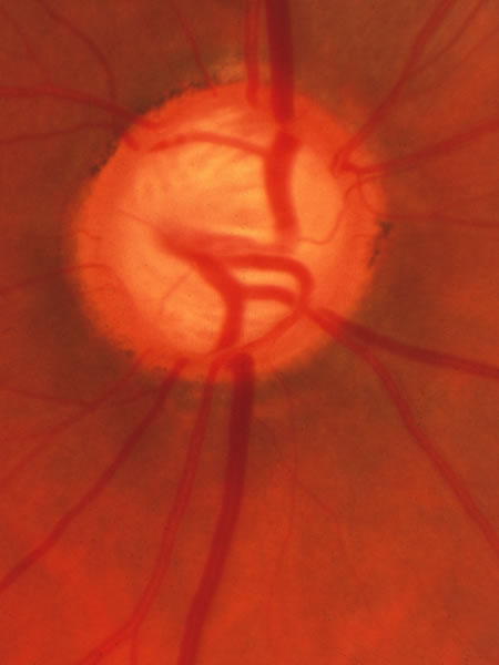
| Fig. 13. Large disc that had a large physiologic cup, on which is superimposed glaucomatous tissue loss. The glaucomatous damage can be recognized by the fact that in the upper and lower sectors the tissue is thinner than nasally and temporally. In particular, careful inspection shows that at the inferior pole in a narrow region where a small venous tributary crosses the margin at the 6:30 position, the cup reaches just about to the disc margin, which is the inner edge of the white stripe that represents the scleral lip. The scleral lip sometimes has a sufficiently pink hue that is mistaken to be remaining neuroretinal tissue in a location where in fact there are not remaining axons. The ongoing glaucomatous process was signified by occasional splinter hemorrhages in this patient. Because the physiologic cup was large initially, the visual loss is strikingly moderate because despite the large cup, only a modest proportion of the original neuroretinal tissue has been lost. |